
Medial lemniscus
(Redirected from Reil's ribbon)
| Medial lemniscus | |
|---|---|
 The sensory tract. (Medial lemniscus labeled at top right.) | |
 Coronal section through mid-brain. ("e" is Portion of medial lemniscus, which runs to the lentiform nucleus and insula. "a’" is also the medial lemniscus.) | |
| Details | |
| Identifiers | |
| Latin | lemniscus medialis |
| NeuroLex ID | birnlex_887 |
| TA98 | A14.1.04.111 A14.1.08.672 A14.1.06.207 |
| TA2 | 5861 |
| FMA | 83675 |
| Anatomical terms of neuroanatomy | |
In neuroanatomy, the medial lemniscus (also known as Reil's band or Reil's ribbon (for German anatomist Johann Christian Reil) is a large ascending bundle of heavily myelinated decussating (crossing-over) second-order axons situated in the medulla oblongata of the brainstem. It consists of decussating internal arcuate fibers (which originate in the nucleus gracilis and nucleus cuneatus). The medial lemniscus is part of the dorsal column–medial lemniscus pathway which conveys fine touch, vibration, and proprioceptive sensory stimuli from sensory receptors to the thalamus. Lesion of the medial lemnisci cause impairment of vibratory and touch sensation.
Anatomy
[edit]Structure
[edit]- At the open medulla (further up the brainstem), the medial lemniscus contains axons from the trigeminal nerve (which supplies the head region), as well as the arms and legs. It sits very close to the midline, at the same orientation of the midline, with head fibres more dorsal (closer to the back), towards the fourth ventricle.
- By mid-pons, the medial lemniscus has rotated. Fibres from the head are medial, fibres from the leg are lateral.[citation needed]
- In the midbrain, it is situated dorsal/posterior to the substantia nigra, and medial to either red nucleus.[1]
- It terminates by synapsing with third-order neurons in the ventral posterolateral nucleus of thalamus[1]
See also
[edit]Additional images
[edit]-
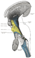 Deep dissection of brain-stem. Lateral view.
 -
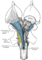 Deep dissection of brain-stem. Ventral view.
 -
 Coronal section of the pons, at its upper part.
 -
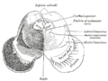 Transverse section of mid-brain at level of inferior colliculi.
 -
 Scheme showing the course of the fibers of the lemniscus; medial lemniscus in blue, lateral in red.
 -
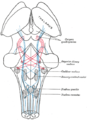
 Horizontal section through the lower part of the pons. The medial lemniscus is labeled #17.
 -
 Tractography showing medial lemniscus


References
[edit]- ^ a b "medial lemniscus - Dictionnaire médical de l'Académie de Médecine". www.academie-medecine.fr. Retrieved 2024-07-27.
- ^ Purves et al, Neuroscience 5th edition, Sinauer Massachusetts, p. 198